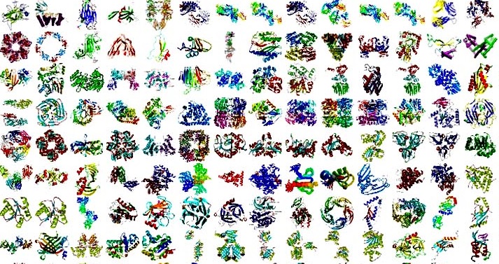
Unravelling the complexity of proteins
 Knowledge of the three-dimensional structures of proteins is
essential for understanding biological processes.
Structures help to explain molecular and biochemical
functions, visualize details of macromolecular interactions, facilitate
understanding of underlying biochemical mechanisms and define biological
concepts.
The human genome and follow-up sequencing projects have
revolutionized biology and medicine; structural genomic programmes have
developed and applied structure-determination pipelines to a wide range of
protein targets, facilitating the visualization of macromolecular interactions
and the understanding of their molecular and biochemical functions.
A paper recently published by Mizianty et al. (2014). Acta Cryst.
D70, 2781-2793; doi:10.1107/S1399004714019427
seeks to address the fundamental question of whether the three-dimensional
structures of all proteins and all functional annotations can be determined
using X-ray crystallography.
The researchers set out to perform the first large scale
analysis of its kind covering all known complete proteomes (the sets of
proteins thought to be expressed by an organism whose genome has been completely
sequenced, as defined by the UniProt Consortium in 2012) and all functional and
localization annotations available in the Gene Ontology for the corresponding
proteins.
The Canadian and US team show that current X-ray
crystallographic knowhow combined with homology modeling can provide structures
for 25% of modelling families (protein clusters for which structural models can
be obtained through homology modelling), with at least one structural model
produced for each Gene Ontology functional annotation. The coverage varies
between super-kingdoms, with 19% for eukaryotes, 35% for bacteria and 49% for
archaea, and with those of viruses following the coverage values of their
hosts. It is shown that the crystallization propensities of proteomes from the
taxonomic super kingdoms are distinct. The use of knowledge-based target
selection is shown to substantially increase the ability to produce X-ray
structures.
Talking to the IUCr Mizianty commented “We believe our
method has helped to advance our understanding of the coverage by X-ray
structures of proteins and complete proteomes on a global scale”.


